InformationPrimary endodontic treatment Endodontic retreatment Special treatments Endodontics step by stepProduct catalogueOrder and payment information Previous special offers Hand instruments Rotary instruments Operating Microscope General instruments Accessoires Filling materials MiscellaneousRemainingReferences Site map Presentations (RECOMMENDABLE!) Downloads Het Kanaal Links Endoplaza Forum MarketPlaza About Endoplaza Instructions for use website
|
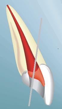
Cleaning and designing the canalsDetails per element11 and 21
Do not forget that a front element is seldom or never perforated to palatinal, which would however be perforated to buccal, especially when a root post should be placed. When a canal cannot be found, you should search further to palatinal. 12 and 22The lateral incisives often have an oval canal (to be observed well with the OPMI). The oval part continues until very close to the apex. It is wise to drill a little bit wider than it would seem normal on the basis of the jamming of the first file. The oval parts can be cleaned until the apical third part with Gates drills or the SX of the Protaper. On the lateral incisive more often than usual an apex resection is done! When you use files< bigger than 25, it is wise to use NiTi files (both hand and Rotary files), because the apex regularly shows quite a curving in the root. In this way you prevent that an own canal is made (ledge) by a tough file. 13 and 23The canine teeth are also oval in the coronary part of the radix. They should be cleaned in the same way as the lateral incisives. 14 and 24It is standard practice that the first premolars have two canals, sometimes even three. Then the two buccal canals find themselves more to distal and mesial. Pay attention to the buccal contour of the element when the buccal canal is not on the expected place. In case of three canals, the latter is less round than usual. Do not file with too big files until the apex because the radices sometimes are very thin. 15 and 25In most cases there is only one canal. Because of the very elongated canal entrance from buccal to palatinal, it often seems to be two canals. Yet only one canal remains when it is cleaned and designed well. The Gates, SX file or a file in the Endo-Eze corner part are fit to clean the complete canal system. 16 and 26
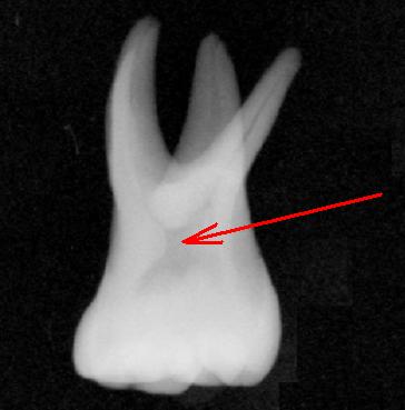
Usually these elements have three roots and four canals. The MB2 canal is often overlooked, but the canal entrance can be found with the OPMI. Nethylene blue colours the entrance and makes it visible. Accessibility to the canal is an indispensable requirement. If the file cannot go further than 2 to 3 mm into the canal, then the dentin wall to mesial should be scraped further. 17 and 27The problems are almost the same as for the 16 and 26. The only difference is that the canals lie closer to each other and more on one single line (MB2 lies more in the direction of palatinal). The percentage of elements with a fourth canal is a little lower here than with the 16 and 26. 32, 31, 41 and 42These elements have regularly two canals or a strong oval canal. If a canal is missing, this is almost always the lingual canal. While opening it, the second canal should be searched until you are sure that this one is not present. The OPMI once again is a great help to find the canals. Do not work with too thick files and be careful when you fill with lateral condensation. With such thin roots, too much pressure in the canals easily leads to a vertical fracture. 33 and 43The coronary part of the canal is often oval. The oval parts can be cleaned with Gates drillsSX files (Protaper), Ultrasone files or the corner part of Endo-Eze with corresponding files. 44, 45, 34 and 35
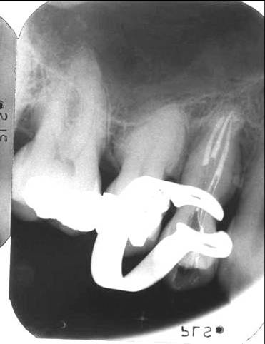
It is almost always a question of oval canals in bucco lingual direction, so that it is difficult to see them on an X-ray. Sometimes the canal seems to be shut at 4 to 6 mm from the apex. Also on the X-ray no canal is visible then. Here it is often a question of a splitting into various canals. If the coronary part has been cleaned sufficiently, with the aid of OPMI you can see if there is a splitting and into what direction the canals go. If between times CA(OH)2 is enclosed, the split canal entrances lighten up beautifully. Also when filling such canals, an OPMI is indispensable (see X-ray).
46 and 36
It is standard practice that 46 and 36 have two roots in which mesially there are always two canals and in which the distal root often has one big oval canal. Apically the two mesial canals have contact regularly (when sucking out one canal the other one empties). Please be careful with rotary files in the mesial canals. The bucco lingual curving apical which sometimes is very strong is almost never visible on the tooth movie. Also the collateral system between MB and ML seizes easily a point of a Rotary file. To remove broken instruments, click here. 47 and 37
Many of the arguments for the 46 and 36 also apply here. With some regularity it is a question of a C-shaped system, which is a difficult incidental circumstance. In fact there is one single canal then, although with the same ease you can make four canals of it. In order to be able to clean the canal system well, an OPMI is indispensable. Then even the deeper and more remote areas of the canal system are clearly visible. Here, all kinds of tools, like SX files (Protaper), Ultrasonic files, Endo-Eze files, Gates drills and hand files have to provide a good final result. Rotary files are a little bit more dangerous here as they easily can get locked in a small niche in the depth. The endo engines which have a spare system can be helpful to prevent a number of fractures. Nevertheless, files can break too!
From this page you can order in our web shop
|